
Muscles
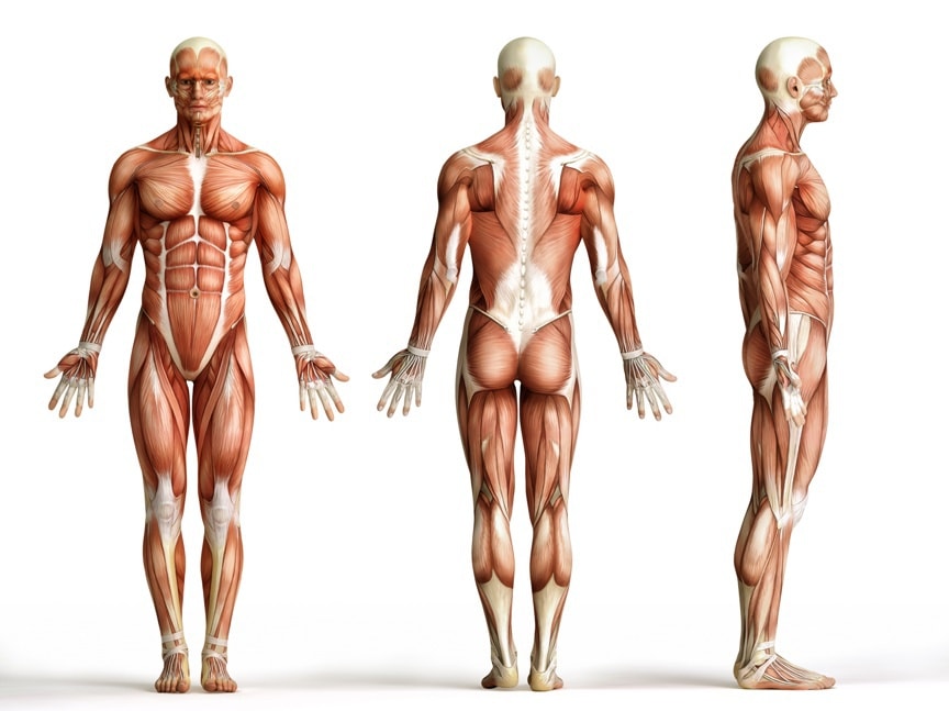
Muscles
The muscles are highly specialized organs that are capable of reducing their length (contracting capacity).
There are the following types of muscles:
- muscle tissue composed of smooth muscle,
- muscles composed of striated muscle fibers subordinate to our will and
- myocardium, consisting of muscle fibers transversely striated independent of our will (from conscious nerve impulses).
The authors provide numerical values for the variable quantity of muscle – about 300 to 500. Skeletal muscle mass ranges from 25 to 35 kg. The colour of muscle is red because of the rich blood supply and because of its own coloration caused by haemoglobin of the muscle. Muscle names are derived from the place of attachment, often they indicate topographic position, the direction of the course, the construction or operation. Muscles, according to the positions, are divided into:
- superficial (musculi cutanei), skin (on the neck, head and face, the palms of the hand) and
- skeletal muscles (musculi skeleti), which are located in the superficial fascia.
With regard to the shape of muscles, we can divide them into:
- long
- wide
- short
Muscles are attached to the surface of the bone; hence there are the initial muscle attachments and the end muscle attachments.
- Every muscle consists of the part referred to the belly (venter), at the end of which there is a second part – connective tendon (tendo).
- The initial part of the muscle is also referred to as the head (caput), the end part – the tail of muscle (cauda).
MICROSCOPIC STRUCTURE
- electron microscopy allows us to distinguish in the histology of muscle the presence of thick filaments of myosin connected temporarily to the thin actin filaments.
- actin and myosin are components of myofilaments and those are the components of myofibrils.
- the grouped muscle fibers are bonded by endomysium.
- primary muscle bundles are connected together by the connective tissue – the inner perimysium (perimysium internum).
- we can also distinguish the external perimysium (perimysium externum), but only in larger muscles
- whole muscle is surrounded by a fibrous membrane – epimysium and this, in turn, by connective membrane – fascia
- the auxiliary organs of muscles include:
- fascia (fasciae)
- synovial bursa (bursae synoviales)
- tendon sheaths (vaginae tendinum)
- navicular bone and muscle blocks (trochleae musculares)
In the process of muscle mechanics, one can distinguish the following muscles: monarticular, biarticular and polyarticular, which in the totality of their operation have an impact on the static and dynamic work, shaping movements and coordination. At the same time, it should be noted that muscle strength is directly dependent on the amount of the muscle fibers contained in it.
MUSCLE UNIT CONTRACTION PHYSIOLOGY
Physiology of muscle contraction comprises:
- slipping of actin filaments, which are attracted by the thickenings of myosin within the muscle band.
- during the contraction there are the alternating processes of releasing and gripping, meaning the alternating insertion of the actin filaments between myosin filaments, there is always one more myosin thickening (strong connection of actin and myosin).
- during rest, there is a process of separation of myosin thickenings from the actin particles.
- muscle tension depends on the content of adenosine triphosphate (ATP), hence during contraction, myosin thickenings in combination with particles of actin, release the degrading enzyme ATP (ATPase ATP>ADP + P + energy) . This reaction is possible due to the presence of calcium ions, which have a significant influence on the release of acetylcholine (A – Ch) into the synaptic cleft and on the opening of channels for sodium cations.
- re – synthesis of ATP occurs through the energy of phosphocreatine in the process of glycolysis and oxidative phosphorylation (ADP + phosphocreatine > ATP + creatine), so it is possible to re-use the high energy compounds of ATP.
- covering the energy demand is variable and depends on the duration of contraction. Energy resources in the form of ATP may be supplied in a short time to about 30 seconds.
- after that time, energy demand participates in the process of glycolysis. Aerobic energy sources have an increasing influence in further work of the muscles.
- we need to remember that a deficiency of calcium ions as well as botulin and excess of magnesium ions clearly influence the difficulties in the process of transmission of nerve impulses and the opening of cation channels.
This information is important as long as we remember about thermal-physiological processes occurring in the muscle unit, which include: heat of activation ( secreted when the muscle is not in operation), – heat of shortening (generated during the internal shortening of the shrinkable elements, occurs also during isometric contraction ), heat of diastole, used up already before the diastole and resting heat, comparable to the initiating heat, provided that there is a sufficient level of oxygen and that the muscle is not tired. To sum up the phase of systole and diastole, one should say:
- During systolic phase, the following processes occur: excitation, depolarization of transverse tubules, release of calcium ions, activation of ATPase of myosin, creation of connections of actin and myosin, slipping of filaments and generation of tension and shortening of the filaments.
- During diastolic phase, the following processes occur: calcium ions pass back to the endoplasmic reticulum, which is connected with a decrease of their value inside the cell, inactivation of ATPase, release of connections of actin and myosin, filaments separation and relaxation of the muscle.
One Response to “Muscles”
Ginnie jaski
Which oil do you remondemd? Can you e-mail me how to studies to become an massage tepartist please such as what to know to the basic know how.